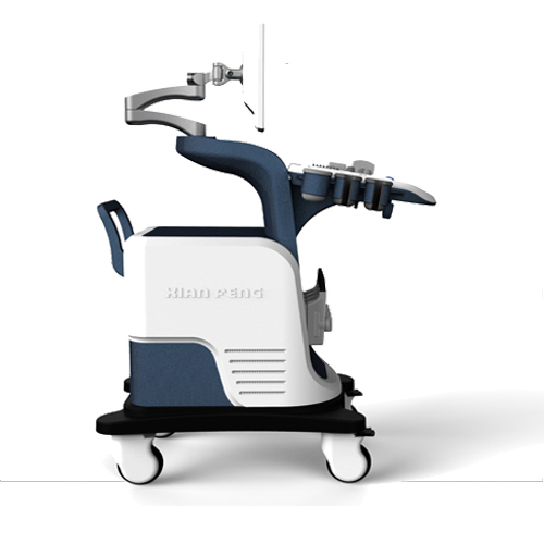


XF7800 color doppler ultrasound system adopts the imaging technology of pioneer high-end
products, with excellent image quality, efficient operation flow and high cost performance,
which can meet the extensive clinical diagnosis needs and create greater value for hospitals.
Humanized ergonomic design
• a 15-inch LCD display that rotates up and down, left and right
• compact fuselage, easy to move
• flexible lifting panel
• two-level backlit keyboard, more convenient to operateIt can meet the needs of clinical diagnosis and improve
the confidence of diagnosis
.• broadband frequency shift harmonic imagingIt can effectively reduce noise and improve contrast resolution
• intelligent speckle noise suppression imagingIntelligent speckle noise suppression technology can intelligently
identify different tissue information in different spatial dimensions, inhibit the display of speckle noise edge
information, and make the image more exquisite.
• intelligent space composite imagingIntelligent spatial composite imaging technology, through
spatial multi-angle deflection scanning, updates echo signals received from different angles
in real time, continuously updates fusion imaging, effectively inhibits random noise and
clutter signals under the premise of guaranteeing the temporal resolution, enhances the
target organization display, and improves the spatial resolution of the image.High resolution
medical liquid crystal display
• flexible adjustment of control panel
Main feature:
*High-definition digital continuous beam former
*Dynamic Frequency Fusion Imaging technology:
*High-definition delay point-by-point dynamic receive focusing:
*Ultra-wideband Imaging technology:
*Adaptive image optimization technology:
*Adaptive angiography
*Adaptive Doppler Imaging technology
*Tissue Harmonic Imaging technology
XF7800 color doppler ultrasound system

Image processing Instruction
Sound beam processing
Image Pre-processing
Total gain: 0~100 adjustable
TGC: 8-segment TGC adjustable
Acoustic output: Low, medium and High 3 levels adjustable
Gray-level: 0-15 levels adjustable
Number of digital channels: 32
Scanning parameters:
XF7800 color doppler ultrasound system

Image Display:
256 levels gray scale display two-dimensional image
Display histogram
Image rotation: left/right, up/down, 90-degree rotation.
Depth range: 3-24 cm (22 levels) each probe has a corresponding depth range
Focus mode: Continuous dynamic focus, dynamic aperture
Dynamic range: ≥120dB (visual, adjustable)
M mode speed: 3-level adjustable
Changing perspectives: 3 kinds of angle adjustable (only applies to convex array)
Display TGC curve
Black-White correlation
Video output: VGA
Measurement/Calculation:
General measurement
B Mode measurement: distance, area (distance measurement method, ellipse distance method, trace method,
line method), circumstance (distance measurement method, ellipse distance method, trace method, line
method), volume (double-plane method, trace-length method, ellipse-length method, diameter method,
ellipse method), angle and ratio.
M Mode measurement: slope, ratio, distance, heart rate and time.
D Mode measurement: Flow Doppler measurement, velocity, acceleration, time, ratio, RI (Resistance Index)
and stroke volume.
Gynecology measurement and analysis
Measurement items include uterus, endometrium, ovary, cervix, and follicle.
Obstetric measurement and analysis
GS(Gestational Sac), CRL(Crown Rump Length), LV(Lumbar Vertebra), BPD(Biparietal Diameter),
OFD(Occipitofrontal Diameter), HC(Head Circumference), TAD(Transverse Abdomen Diameter), LVW(Width
of posterior horn of lateral ventricle), HW(Hemispheres Width), TCD(Transverse Cerebellar Diameter), IOD
(Orbita Insider Diameter), OOD(Outer Orbita Diameter), BD(Binocular Diameter), APTD(Thorax Anterior-Posterior
Diameter), TTD(Thorax Transverse Diameter), AC(Abdomen Circumference), APD(Abdominal Anterior-Posterior
Diameter), FTA(Fetal Trunk Abdominal area), HL(Humerus Length), ULNA(Ulna length), RAD(Radius length),
FL(Femur Length), TIB (Tibia length), FIB (Fibula length), APTDxTTD, CLAV(Clavicle length), Hip dysplasia {Hip
angle measurement}etc, can calculation gestational age, fetal weight, estimated delivery date, Chinese
population formula, Fetal physiology score.
Urology measurement and analysis
Prostate volume, Bladder volume, Residual Urine Volume, Prostate Transition Zone, Hip angle measurement
and evaluation (newborn hip dislocation diagnosis), section measurement (V-Slice)
Cardiology measurement and analysis
Measurement items in B Mode, B/B Mode, CFM Mode, PDI Mode:
Mitral valve: E-point septal separation (EPSS), Left Ventricular Outflow Tract (LVOT) Diameter, Mitral valve
area, Mitral valve diameter
Aorta/Left atrium: LVOT Diameter, Aorta cusp space (AoCS), Aorta/Left atrium
Aortic Valve: LVOT Diameter, Aorta Valve Area
Left Ventricle Measurement: Diastole, Systole
Left Ventricle function
LV Mass
l Measurement items in M, /B/M Mode:
Aorta/Left atrium: LVOT Diameter, Aorta cusp space (AoCS), Left Ventricle Ejection Time, Left Ventricular
Pre-ejection Period (LVPEP), Aorta/Left atrium
Mitral valve: E wave amplitude, A wave amplitude, Diastole excursion, DE amplitude, E-point septal separation
(EPSS), EF slope
Left Ventricle Measurement: Diastole, Systole
Left Ventricle function
l Measurement items in PW Mode:
Left Ventricular Outflow Tract (LVOT):LVOT, Aorta Valve Area
Mitral valve: Ratio E/A, Velocity integral (VTI), Mitral valve Area, Isovolumic Relaxation Time (IVRT), Heart Rate
Aortic Valve Systole: Velocity integral (VTI), Acceleration, Aorta Valve Area, Heart Rate
Tricuspid valves: Ratio E/A, Velocity integral (VTI), Tricuspid valve area, Heart Rate
Pulmonary Valve: Velocity integral (VTI), acceleration
Pulmonary veins: Systole/Diastole, A. Rev Velocity, A. Rev Duration
Tissue Doppler: Ratio E/A, Isovolumic Relaxation Time (IVRT), Acceleration time, Deceleration time
Left-to-right shunt ratio (Qp/Qs): Pulmonary Valve, Aortic Valve
Small parts and Peripheral Vessel measurement and analysis
Main measurement and analysis: vessel sectional area, heart rate, stroke volume, Unit time flow, Ejection
time, Stenosis ratio, MFV (Mean Flow Velocity), RI (Resistance Index) and PI (Pulsatility Index).
Measurement report
Obstetric measurement report, gynecology measurement report, cardiology measurement report, urology
measurement report and other measurement reports Automatically store measurement results and generate report.
Marks
More than or equal to 95 kinds of body marks with probe position, which can be quickly selected by intuitive
and detailed body mark interface Text mark can preset text content Arrow mark supports multiple arrow marks
and adjustable direction Patient data: Medical record management, report inquiry and printing, image video
output( HDD ,USB, Optional DVD-RW),built-in ultrasound workstation;Reporting system: automatic report
generation system, and can be full screen characters in both Chinese and English editor;
Standard configuration:
Host .convex probe.linear probe.trans-vaginal probe. one each;
Image storage: Image storage, video storage, cine loop, disk storage capacity≥120G;
Output interface:SR323,USB,DICOM interface;
Displayer: 15 or 17 inch LCD color display;
Running hours: ≥8h;
Input power: ≤320V;
Host weight: about 80 kg;
Host appearance size: 950 ×520× 1260(length × width × height) (mm3).