1. Application
1.1. Urological calculi
1.2. Urological operation table and X-ray fluoroscopy
2. Working condition
2.1. Environmental temperature 15~35℃
2.2. Relative humid 45~85%
2.3. Atmospheric pressure86~106kPa
2.4. Power requirements: AC 220V 50/60±1Hz, PW≤3.5kW
2.5. Water requirements: Dematerialized water less than 20L once
2.6. Room requirements:
2.6.1. Main room ≥3m×3m×2.5m
2.6.2. Auxiliary room ≥2m×4m
2.6.3. X-ray proof of the main room
3. Shock wave generator: Electromagnetic
3.1. Discharging range: 10~16kV
3.2. Parameter of second focus of shock wave
3.2.1. Shock wave front≤ 0.5mS
3.2.2. Shock wave width≤ 1mS
3.2.3. Focal spot area: 7.5×7.5×40 mm (standard)
3.2.4. Peak value of second focus of shock wave: 20~50mPa
3.3. Length of second focus: ≥140mm
3.4 Shock wave generator level variation: 45°~ 25°
3.5 Focus as the center of a circle, shock wave generator can rotate 180°
4. Localization system
4.1. X-ray localization (C arm & X-ray double localization)
4.1.1. X-ray module of 3.5kw/ 40kHZ
4.1.2. X-ray high-voltage generator voltage range: 40-110kv
4.1.3 X-ray high-voltage generator current range: 0-5mA
4.1.4 Tube focus size: 0.6/1.5mm
4.2. B-ultrasound localization (Optional)
4.2.1. Manipulator rotate 360° round shock wave source.
4.2.2. Manipulator and shock wave centerline: 45°~ 90°
4.2.3.The localization error may be caused by manipulator changes the probe axis relative to the focus is within ±2mm
5. Image system
5.1. High resolution 9" image intensifier
5.2. High definition CCD camera
5.3. High definition and multifunction digital monitor
5.4. Image de-noise and last image hold function
5.5. X-ray image definition ≥12 line/cm
6. Operating system
6.1. Compartment and bedside operating system
6.2. Wireless remote control operating system
6.3. PLC central control system
6.4. Liquid crystal monitor operating information
6.5. Shock wave triggering frequency: 45~120/mi
6.6. Triggering mode: manual operation, continuous operation and electrocardio synchronousness
6.7. Compartment talk back equipment
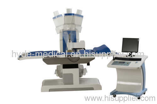
7. The main unit and therapy table
7.1. C arm angle of rotation: ±30
7.1. 3-D mechanical movement: X≤100mm, Y≤100mm,Z≤250mm
7.2. Minimum adjustment of 3-D movement of the table≤1mm
7.3. Maximum capacity of the table: 135kg
7.4. The localization error may be caused by C arm and wave source movement relative to the second focus within ±2mm
8. Water treatment system
8.1. Water automatic degassing and cycle
8.2. Water automatic thermostatic apparatus
8.3. Overtemperature alarm and protective device
9. Image workstation
9.1. Computer hardware
9.2. Hyde image software can support medical record, image processing saving and network sharing, etc.
9.3. Laser printer
10. Major components' volume and weight
10.1. Host
10.2. Therapy table
10.3. Console
10.4. Accessory case
Ⅲ Standard layout of equipment(reference)
1.Big and small C arm hosts
2.Therapy table
3.Console and bedside control box, remote control box
4.Electromagnetic shock wave source (electromagnetic tube, capacitor and cusion)
5.High frequency X-ray inverter (40KHz, 3.5Kw, static anode)
6.High frequency high voltage generator and X-ray tube (all-in-one)
7.High resolution 9inch image intensifier
8.High definition CCD camera (400 thousand pixel)
9.Digital multifunctional monitor (14″)
10.Computer hardware (include 17″ liquid crystal monitor) and image workstation software
11.Talkback unit
12.Enclosure
Ⅳ Changeable layout and performance
1.Host can use connecting lever host ( wave source can be moved from the bed)
2.Choose electrohydraulic wave source or add electrohydraulic wave source interchanging
3.Use 6″ intensifier
4.Choose 5Kw high frequency X-ray inverter ( rotating anode)
5.Use power frequency X-ray device
6.PLC central control system can be changed by computer control system
6.1. Operation interface uses widescreen touch screen and mouse control
6.2. X-ray pulse exposure and automatic KV brightness control
6.3. Ultrasound localization automatic follow fragments calculi
Remarks: The functions, parameters and configuration mentioned above are just for conference, and they would be changed according to customer's specific requirements and the final agreements or contracts with the customer.